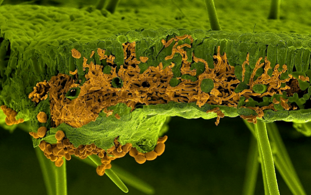
Scanning electron microscopy picture of a soybean leaf infected by the rust fungus Phakopsora pachyrhizi. The leaf and the fungus were artificially painted in green and in orange, respectively. The section shows invading infection hyphae of the fungus inside the leaf mesophyll, whereas the spores are visible below the leaf breaking through the lower epidermis (U. Steffens, Bayer Crop Science)