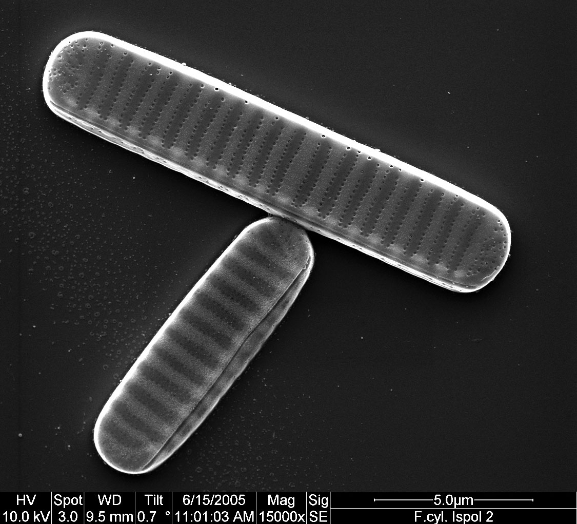
Scanning electron micrograph of two cells of Fragilariopsis cylindrus. Shown are two silica shells (Frustules) in valve view. Magnification: 15,000X; scale bar: 5 μm (Image credit: Gerhard S. Dieckmann)
Scanning electron micrograph of two cells of Fragilariopsis cylindrus. Shown are two silica shells (Frustules) in valve view. Magnification: 15,000X; scale bar: 5 μm (Image credit: Gerhard S. Dieckmann)