Archive

  • Visit JGI.DOE.GOV
All JGI Features

August 11, 2014

Cryptococcus neoformans giant cell SEM

Cryptococcus neoformans giant cell SEM

Cryptococcus neoformans giant cell.

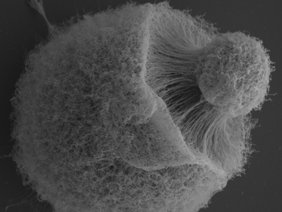
Scanning electron microscopy image of a Cryptococcus neoformans giant cell isolated from the lung of an infected mouse. The image illustrates one of the most characteristic phenotypic features of giant cells, which is the high density of polysaccharide fibers present in the capsule. This structure presents as a highly cross-linked net, which is accumulated as a very compact layer around the cell body (see Zaragoza et al., doi:10.1371/journal.ppat.1000945).

Image Credit: Oscar Zaragoza, Instituto de Salud Carlos III, Spain, and Albert Einstein College of Medicine, United States of America

More from the JGI archives:

  • Software Tools
  • Science Highlights
  • News Releases
  • Blog
  • User Proposals
  • 2018-24 Strategic Plan
  • Progress Reports
  • Historical Primers
  • Legacy Projects
  • Past Events
  • JGI.DOE.GOV
  • Disclaimer
  • Accessibility / Section 508
Lawrence Berkeley National Lab Biosciences Area
A project of the US Department of Energy, Office of Science

JGI is a DOE Office of Science User Facility managed by Lawrence Berkeley National Laboratory

© 1997-2025 The Regents of the University of California